Using Image Analysis for the Detection and Evaluation of Wrinkles
Typically photographs taken during a clinical study are used to document visible changes in skin conditions and for marketing purposes. By using Stephens’ image analysis, which can detect and quantify changes in an objective manner, you can maximize the use of your images by collecting additional data from them.
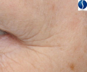
Our wrinkle image analysis, SWIRL (Stephens Wrinkle Imaging using Raking Light), will provide you with the number, length, area and depth of the wrinkles on multiple facial areas.
 We provide image analysis on any clinical photographs, whether they are taken at one of our sites or at a different site. The key is to have high quality images with the correct lighting mode. We are happy to assist you to determine the correct lighting mode and answer any questions on how to obtain high quality images for the analysis.
At the most recent ISBS meeting in Connecticut, Dr. Lily Jiang presented Stephens’ poster entitled “Quantitative analysis of multiple photoaging features using image analysis of digital photographs” and won best poster! SWIRL was also published in the June 2013 issue of the peer-reviewed journal, Skin Research and Technology.
Stephens also provides image analysis for a variety of other skin parameters. A full listing is located here.
Maximize the use of your images and contact Dr. Lily Jiang to use Stephens’ image analysis and to learn more about our image analysis services.
Want to participate in a study?
We are always seeking panelists for clinical trials. Participating in a study is a great way to earn money, meet fun people, and fill your day with meaningful work! Choose your preferred clinical study location below to learn more.