Image Analysis
SGS continually works to innovate in developing processes for advanced evaluation and reporting techniques in clinical studies.
Historically, photographs of subjects were only used to document clinical changes, but with new innovative methodologies SGS has developed image analysis macros in conjunction with Image Pro™, and data can be collected and evaluated from your clinical trial photographs. Our image analysis will provide you with a new, powerful and objective way to detect and quantify changes in various skin parameters.
Our image analysis method, SWIRL (Stephens Wrinkle Imaging using Raking Light), has been published in the June 2013 issue of the peer-reviewed journal, Skin Research and Technology. See below for more details on the method.
In addition to the wrinkle analysis, we can analyze your images for the features listed below. If a feature you are interested in is not listed below, please let us know and we would collaborate with you to develop it.
- Pigmentation
- Pores
- Erythema
- Skin Tone
- Oily Skin
- Dry Skin
- Facial Cleansing (example: % makeup removed)
- Stratum Corneum Turn over
- Skin Roughness
- Fingernail Growth
- Hair Growth
- Silicone Replicas (Wrinkles and Roughness)
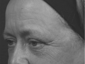
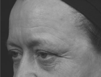
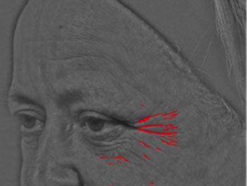
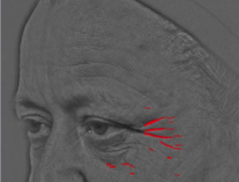
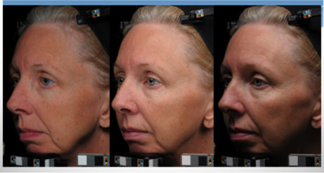
SWIRL (Stephens Wrinkle Imaging Using Raking Light)
SGS began developing the method in response to the increasing demand for an objective and quantitative assessment of facial wrinkles before and after treatment from prescription products, cosmetic products or aesthetic procedures. The SWIRL method analyzes the wrinkle severity at multiple areas on the face, such as the crow’s feet, under eye, forehead, and upper lip areas. Five parameters are quantified and reported including the number, length, width, area, and depth of wrinkles. This method has been validated through clinical studies and demonstrates excellent correlation with clinical grading.
